Glossary (sequenced by the first letter)
Accommodation: Accommodation is a process of optical power changes of eyes for keeping a legible vision when the range between the observed and the observer shifts. The dynamic process of a lens’ morphological transformation is caused by the changes in the radius of the ciliary muscle (Helmholtz 1909), during which the curving covering diameters of the lens on both sides reduce, resulting in a raising power of refraction (Glasser 2006), then the lens will be accommodated (Figure 1).
Figure 1. Contraction of the ciliary muscle results to an accommodation (Land 2005)
Blind spot: The blind spot is a small region in the field of vision correlating to the location of the optic disc (Figure 2) without image detection (Gamm, Albert & Daniel 2020), from which is the cranial nerve II penetrates so that there are no photoreceptors in the optic disc (Ramachandran 1992).
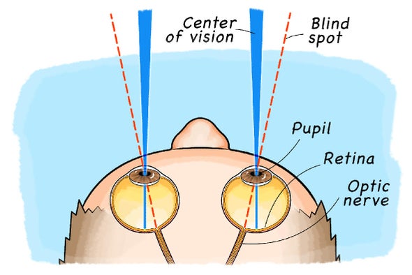
Figure 2. Location of blind spot (Lohner 2020).
Cones and rods: Cones and rods are the two kinds of photoreceptors (Figure 3) in the retina of the vertebrate retina (Schwiegerling 2004). The cones show responsibility to the bright light and colour sensitivity while cones are responsible for dim light (Schultze M 1866).
Figure 3. Differentiation between Cones and Rods (Ingram et al. 2016)
Directionally selective ganglion cells: Directionally selective ganglion cells are a group of retinal ganglion cells that can detect motions in different directions. They can respond actively to a movement from the chosen direction with a high postsynaptic potential after an inhibitory spike (Borst 2011) while active infrequently in response to opposite-direction movement of the same item (Barlow 1963).
Fovea Centralis: Fovea Centralis is a tiny fossa in the centre of eyes (Figure 4) composed of cones densely located in the middle of the macula lutea (Iwasaki & Inomata 1986). Foveal vision is functioned by this area and is responsible for distinct item characteristics, such as form and location (Lettvin 1976).
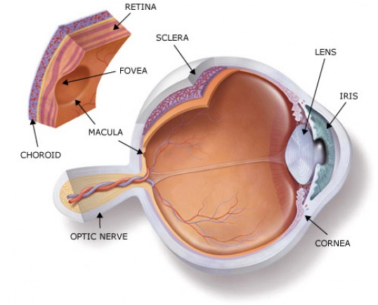
Figure 4. Fovea centralis anatomy in human eyes
Photopigment: Photopigment is a transmembrane protein held inside photoreceptors functioning for light perception situated in plates of film in the external fragment of a pole or cone (Fu 2010). The purpose is to absorb incident light and afterwards produce a cascade reaction to change the electrical characters of the photoreceptors, in which case trigger the release of glutamate (Hart 2009).
Photoreception: Photoreception is a process that photoreceptors for instance cones and rods retain light waves which are captured by eyes then convert the electromagnetic signs into electrochemical signs which are then shipped off the cerebrum for visual handling invertebrate animals (Foster et al. 1991). The whole process converts light stimulus to nerve impulse at last with help from specialised photoreceptors in the retina.
Phototransduction: Phototransduction is an interaction by which electromagnetic energy is changed over into electrical chemical energy in cone cells, rod cells and retinal photosensitive ganglion cells in the eyes (Kusakabe et al. 2009). This process includes a series of primary biochemical and physiological actions, which is also known as “Wald’s Visual Cycle” (Figure 5).
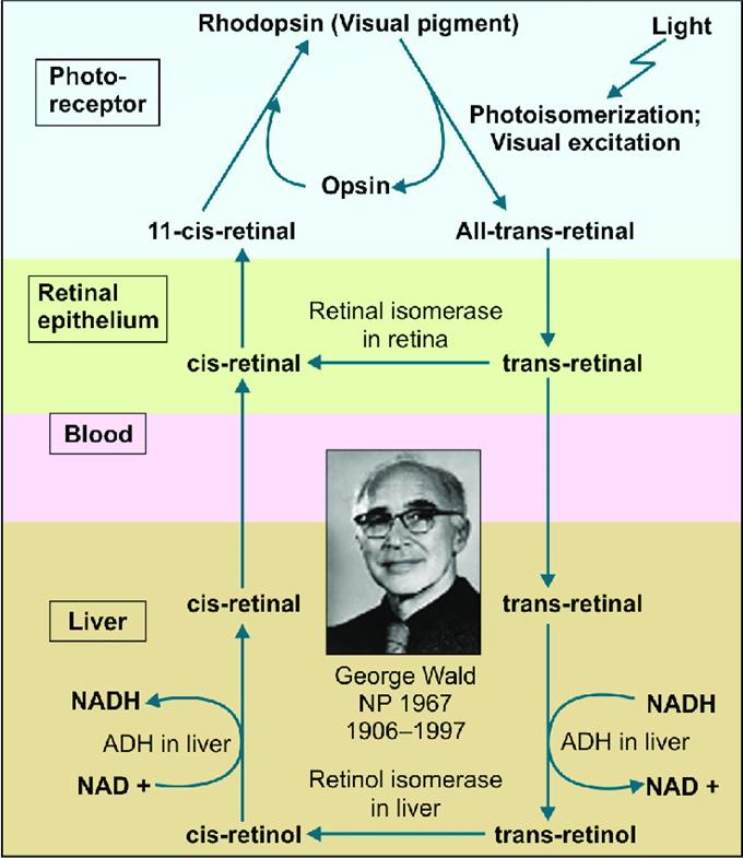
Figure 5. Wald’s Visual Cycle (Vasudevan et al. 2017)
Retina: The retina is the inverted tissue component toward the rear of the eyes (Figure 6) that detects light after cones and rods and sends processed information to the cerebrum. It is a photoreceptor and glial cells layer that catches approaching photons and sends them along neuronal pathways as both electrical and synthetic signs (Nguyen et al. 2021).
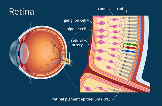
Figure 6. Retina anatomy (Heiting 2019)
Rhodopsin: Rhodopsin is a kind of light-sensitive G-protein coupled receptor protein (GPCR) (Figure 7) (Lenaan et al. 2020) functioning during vision Wald’s visual cycle. As a photo switchable opsin, one kind of photopigment found in retinal rods, it can be activated in a very low light environment (Litmann 1996).
Figure 7. 3D view of rhodopsin model (1F88)
References:
Helmholtz, H. & Southall, J. P. C. (1962). Helmholtz’s treatise on physiological optics. New York: Dover Publications.
Land, M. (2015). Focusing by shape change in the lens of the eye: a commentary on Young (1801) ‘On the mechanism of the eye’. Philosophical transactions of the Royal Society of London. Series B, Biological sciences, 370(1666), 20140308. https://doi.org/10.1098/rstb.2014.0308
Glasser, A. (2006). Restoration of accommodation. Current opinion in ophthalmology, 17(1), 12–18. https://doi.org/10.1097/01.icu.0000193069.32369.e1
Gamm, D. M. Albert, and Daniel M. (2020). blind spot. Encyclopedia Britannica. https://www.britannica.com/science/blind-spot
Ramachandran, V. S. (1992). Blind spots. Scientific American, 266(5), 86–91. https://doi.org/10.1038/scientificamerican0592-86
Lohner, S., (2020). Find Your Blind Spot! Scientific American.
Schwiegerling, J. Field Guide to Visual and Ophthalmic Optics, SPIE Press, Bellingham, WA (2004)
Schultze M (1866). Zur Anatomie und Physiologie der Retina.Archiv f ̈ur mikroskopische Anatomie2, 175–286
Ingram, N. T., Sampath, A. P. & Fain, G. L. (2016). Why are rods more sensitive than cones? The journal of physiology, 594 (19), s. 5415–5426. doi:10.1113/jp272556
Borst A, Euler T (2011). “Seeing things in motion: models, circuits, and mechanisms”. Neuron. 71 (6): 974–94.
Barlow, H. B. & Hill, R. M. (1963). Selective sensitivity to direction of movement in ganglion cells of rabbit retina. Science 139, 412–414.
Iwasaki, M., & Inomata, H. (1986). Relation between superficial capillaries and foveal structures in the human retina. Investigative Ophthalmology & Visual Science, 27(12), 1698-1705.
Lettvin J. Y. (1976). On seeing sidelong. The Sciences, 16(4), 10–20, 10.1002/j.2326-1951.1976. tb01231.x
Myeyepage.weebly.com. 2021. myeyepage.weebly.com - Fovea Centralis.
Hart, N. S. (2009). Photopigments. In M. D. Binder, N. Hirokawa, & U. Windhorst (Eds.), Encyclopedia of Neuroscience (pp. 3148-3151). Berlin, Heidelberg: Springer Berlin Heidelberg.
Foster, R.G.; Provencio, I.; Hudson, D.; Fiske, S.; Grip, W.; Menaker, M. (1991). “Circadian photoreception in the retinally degenerate mouse (rd/rd)”. Journal of Comparative Physiology A. 169 (1): 39–50. doi:10.1007/BF00198171. PMID 1941717. S2CID 1124159.
Fu, Y. (2010). Phototransduction in Rods and Cones. In H. Kolb (Eds.) et. al., Webvision: The Organization of the Retina and Visual System. University of Utah Health Sciences Center.
Nguyen, K. H., Patel, B. C., & Tadi, P. (2021). Anatomy, Head and Neck, Eye Retina. In StatPearls. StatPearls Publishing.
Heiting, G., (2019). How the Retina Works - Detailed Illustration. All About Vision.
Kusakabe, T. G., Takimoto, N., Jin, M., & Tsuda, M. (2009). Evolution and the origin of the visual retinoid cycle in vertebrates. Philosophical Transactions of the Royal Society B: Biological Sciences, 364(1531), 2897-2910. doi:10.1098/rstb.2009.0043
Vasudevan, Damodaran & S, Sreekumari & Vaidyanathan, Kannan. (2017). Chapter-15 Fat Soluble Vitamins. 10.5005/jp/books/13106_16.
Lenahan, C., Sanghavi, R., Huang, L., & Zhang, J. H. (2020). Rhodopsin: A Potential Biomarker for Neurodegenerative Diseases. Frontiers in neuroscience, 14, 326. https://doi.org/10.3389/fnins.2020.00326
Litmann BJ, Mitchell DC (1996). “Rhodopsin structure and function”. In Lee AG (ed.). Rhodopsin and G-Protein Linked Receptors, Part A (Vol 2, 1996) (2 Vol Set). Greenwich, Conn: JAI Press. pp. 1–32.